Our Services

High MRI FAQ

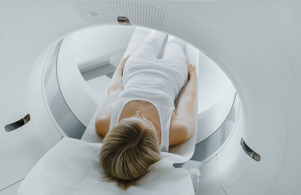
During an MRI scan, patients will lie down on a table that slides into a large, cylindrical magnet. The machine uses strong magnetic fields to create detailed images of your body. It is extremely important to remain still during the scan, as movement can blur the images. The technologist may provide padding or cushions to help you stay in the correct position and remain comfortable.

Throughout the scan, you will be able to communicate with the MRI technologist via an intercom system. They will give you instructions and updates on the progress of the scan. If you feel uncomfortable or experience any issues, you can alert them immediately.

The procedure is painless, but you may hear loud noises during the procedure. Earplugs or headphones are usually provided to help reduce the noise. Once the scan is complete, the table will slide out of the MRI machine. Patients can resume normal activities unless instructed otherwise by their healthcare provider.

MRI is a non-invasive and generally safe imaging technique with minimal risks. Unlike X-rays or CT scans, MRI does not use ionizing radiation. However, individuals with certain implants, medical conditions, or medical devices may need to avoid MRI scans or take special precautions. Always inform your healthcare provider of any metal implants or health conditions before the procedure.

Preparation for an MRI is typically straightforward. Patients can eat and drink normally, and continue taking medications unless otherwise advised by the healthcare provider. You may be asked to remove any metal objects, such as jewelry, clothing with metal fastenings, or other personal items that could interfere with the magnetic field.

All personal items will be secured outside of the exam room. We ask that you avoid wearing makeup or applying other skin care treatments before your procedure. It’s important to follow any specific instructions provided by your healthcare provider or the imaging center to ensure accurate results and a smooth procedure.

Philips Achieva 1.5T High Field MRI

The Philips Achieva 1.5T High-Field MRI is an advanced imaging technology that provides exceptional clarity and detail for diagnosing a wide range of medical conditions. This system allows clear visibility of soft tissues, organs, and structures, making it an invaluable tool for identifying abnormalities, monitoring diseases, and planning treatment strategies.

Designed with comfort in mind, the 1.5T High-Field MRI provides detailed imagery to help physicians with early detection and diagnosis – leading to better outcomes and more personalized care.

Fuji Velocity 1.2T
High Field Open MRI

The Fuji Velocity 1.2T High-Field Open MRI combines advanced imaging technology with the accessibility of an open MRI system. The design of the MRI machine features a more open configuration compared to traditional closed MRI systems, which helps reduce feelings of claustrophobia and provides a more comfortable experience for patients. This is particularly beneficial for individuals who may have difficulty tolerating the confined space of a conventional MRI machine.

The combination of strength and an open design ensures that patients receive comprehensive diagnostic information while experiencing less discomfort and anxiety during the procedure.

Open MRI FAQ

The main difference is the design. Open MRI features an open configuration that reduces the feeling of confinement, making it more comfortable for patients who are anxious about enclosed spaces. Despite its open design, this system consistently provides clear and reliable imagery. The specialized framework also makes it easier to accommodate larger patients and those with mobility issues.

During the scan, patients lie on a table that slides into the Open MRI machine. Unlike traditional MRIs, you will not be fully enclosed to help alleviate feelings of claustrophobia. The machine will produce loud noises, which can be minimized with earplugs or headphones. It’s important to stay as still as possible to ensure clear images. The procedure is typically quick and painless, and you will be able to communicate with the technologist throughout the scan.

Preparation is generally straightforward. You will need to remove any metal objects, such as jewelry, clothing with metal parts, or other items that could interfere with the magnetic field. Talk with your healthcare provider about any medical implants, devices, or health conditions that might affect the MRI. Certain conditions or devices make patients ineligible for an MRI. Follow any specific instructions provided by the imaging center to ensure the best possible results and a smooth scanning experience.

MR Arthrogram FAQ

The procedure begins with the injection of a contrast agent into the joint space, which is guided by fluoroscopy (live X-ray) or ultrasound to ensure accurate placement. Once the contrast agent is injected, imaging is performed using MRI to capture detailed pictures of the joint. The entire process usually takes 30 to 60 minutes and is performed on an outpatient basis.

MR Arthrograms offer several advantages, including improved visualization of joint structures and higher-resolution imagery. This procedure is particularly useful for diagnosing joint issues, cartilage tears, ligament injuries, and subtle joint abnormalities. The extremely detailed imaging helps physicians make more accurate diagnoses and develop tailored treatment plans specifically for the patient’s condition.

Before the procedure, inform your healthcare provider of any medical conditions or metal implants. Certain conditions and medical implant devices make a patient ineligible for MRI.

During the procedure, patients lie on a platform that slides into the MRI machine. A fine needle is inserted into the joint to inject the contrast agent, and the patient is lowered into the MRI. The machine will produce loud noises, which can be minimized with earplugs or headphones provided by the facility. It’s important to stay as still as possible to ensure clear images. The procedure is typically quick and painless, and you will be able to communicate with the technologist throughout the scan.

After the procedure, some patients experience some discomfort, swelling, or bruising at the injection site – which typically resolves within a few days. Most patients can resume normal activities shortly after the procedure, but the physician will provide specific aftercare instructions.

While MR Arthrograms are generally safe, they do come with some potential risks and side effects. These may include infection at the injection site, allergic reactions to the contrast agent, or temporary discomfort and swelling. In rare cases, there may be joint irritation or bleeding.

It is important to discuss any concerns with your healthcare provider beforehand and follow their post-procedure care instructions to minimize risks.

MR Arthrogram

An MR Arthrogram is a specialized diagnostic imaging technique to visualize the internal structures of a joint, like cartilage, ligaments, tendons, and bone. It involves the injection of a medical-grade dye directly into the joint space. This contrast agent allows for a more detailed and clearer view of the joint’s anatomy to see any irregularities, tears, impingements, or degenerative changes.

This advanced imaging tool is valuable for diagnosing complex joint issues that are not easily seen with standard imaging techniques. By integrating MR Arthrograms into the diagnostic toolkit, we can plan and guide treatment, ultimately leading to more effective and individualized patient care.

Get a Clearer Picture of your Health.

Get a Clearer Picture
of your Health.

Contact
Us

If you need to schedule an appointment, inquire about our procedures, or get more information on how we can assist with your health needs, our friendly and knowledgeable staff is ready to assist you! Reach out to us via phone, by visiting our office, or by completing the contact form below. Get picture-perfect diagnostic imaging with Open Air MRI of Lake Charles, today!
Name(Required)